

When you look at a vial of blood drawn during a standard medical check-up, it might just look like a uniform red liquid. However, once that sample undergoes processing in a laboratory, it reveals two distinct components: the cellular elements and the fluid fraction. Understanding what is the difference between serum and plasma is essential for anyone interested in clinical diagnostics, hematology, or general physiology.
While both are the liquid portions of blood that remain after removing red cells, white cells, and platelets, they are not identical. The primary distinction lies in the presence or absence of clotting proteins, a difference that dictates how each substance is handled, processed, and utilized in a clinical setting.
To grasp the distinction, one must first look at the biological process of blood coagulation. Plasma is the liquid component of blood that is obtained when the blood is prevented from clotting.
Conversely, serum is the liquid that remains after the blood has been allowed to clot. This might seem like a subtle procedural shift, but it fundamentally alters the biochemical composition of the fluid, making the choice between them a critical decision for laboratory technicians and pathologists depending on the specific diagnostic test being performed.
The Biological Composition of Blood Plasma
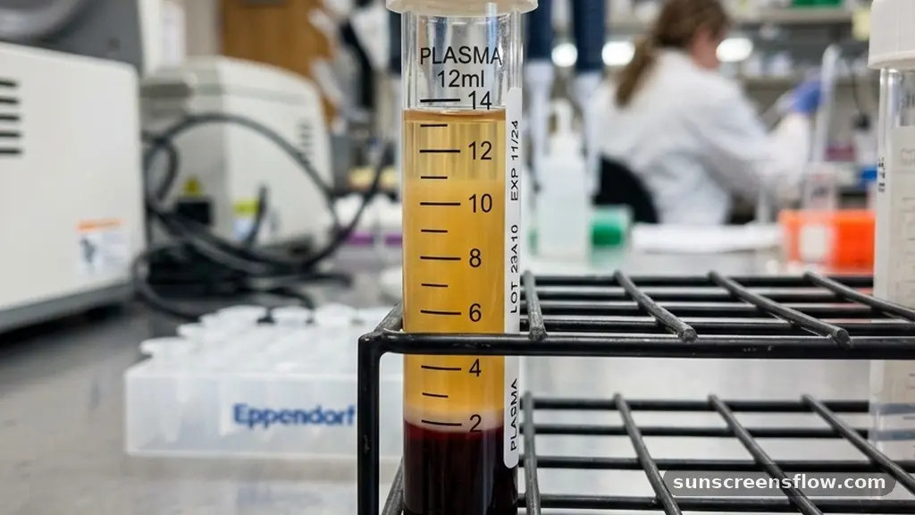
Blood plasma represents the straw-colored, liquid portion of blood that accounts for approximately 55% of total blood volume. It is a complex solution composed primarily of water, but it is also rich in dissolved proteins, glucose, mineral ions, hormones, carbon dioxide, and platelets. Because plasma is collected using anticoagulants—substances that prevent the activation of the coagulation cascade—it retains all the soluble proteins that are otherwise consumed during the formation of a blood clot.
Crucially, plasma contains fibrinogen, a high-molecular-weight protein that is converted into fibrin threads during the clotting process. In a living, circulating system, fibrinogen is essential for maintaining homeostasis and preventing excessive blood loss. When a lab collects plasma, they are essentially capturing a “snapshot” of the blood’s chemical environment as it existed inside the veins.
Because the blood was never allowed to clot, the levels of various proteins and electrolytes remain closer to their physiological state in the body. This makes plasma an invaluable resource for emergency medicine, where rapid analysis of clotting factors or specific proteins is required to save lives.
The Formation and Nature of Serum
Serum is defined as the liquid that remains after the blood has been allowed to undergo full coagulation. When blood is drawn via venipuncture and placed into a standard tube without any anticoagulants, it naturally begins to clot.
During this period, platelet activation occurs, and the coagulation cascade is triggered, leading to the conversion of fibrinogen into solid fibrin. This fibrin mesh traps the cellular components of the blood, forming a solid clot.
As the clot retracts over time, it squeezes out a clear, yellowish fluid: the serum. Because the clotting process has already consumed the clotting factors—specifically fibrinogen and certain enzymes like thrombin—serum is essentially plasma minus these specific proteins. This lack of fibrinogen is the defining characteristic of serum.
While this might seem like a loss of data, it is actually a benefit for many types of diagnostic testing. Because serum lacks the fibrinogen that could potentially interfere with automated laboratory equipment or create “fibrin strands” that clog sensitive machinery, it is often the preferred medium for many routine chemistry panels.
The Role of Centrifugation in Separation
Whether you are working with serum or plasma, the process of separating the liquid from the solid cells involves a centrifuge. A centrifuge is a laboratory device that spins the blood sample at high speeds, utilizing centrifugal force to push the heavier cellular components—red blood cells, white blood cells, and platelets—to the bottom of the tube. The clear liquid that sits on top of this cell pellet is referred to as the supernatant.
For plasma, the blood is collected in a tube containing an anticoagulant and then centrifuged immediately. Because the blood was prevented from clotting, the supernatant is plasma. For serum, the blood is allowed to sit for 30 to 60 minutes to ensure a solid clot forms before it is centrifuged.
The resulting supernatant is serum. The time taken for the clot to form is a significant factor in laboratory workflow.
Using a serum separator tube, which contains a gel that physically separates the clot from the serum during spinning, helps ensure that the serum remains pure and free of cellular debris. This mechanical separation is vital for maintaining the integrity of the sample, as leaving serum in contact with the clot for too long can alter the concentration of certain analytes due to metabolic activity within the trapped cells.
Anticoagulants and Their Impact on Sample Integrity
The chemical environment of a blood sample is heavily influenced by the anticoagulants used during collection. These substances work by binding to calcium or inhibiting specific enzymes in the clotting cascade, effectively “freezing” the blood in its liquid state. Common anticoagulants include:
- EDTA: Widely used for hematology studies because it preserves the morphology of blood cells exceptionally well.
- Heparin: Often used for chemistry panels because it does not interfere with many electrolyte assays.
- Sodium citrate: The standard for coagulation studies because it is easily reversible, allowing the lab to restart the clotting process under controlled conditions.
When you choose a tube for blood collection, you are essentially choosing the final state of your sample. If you use a tube with EDTA, you are creating plasma. If you use a plain tube with no additives, you are creating serum.
It is vital for clinicians to understand that using the wrong tube can lead to skewed results. For example, if a test requires the measurement of calcium levels, using an anticoagulant that chelates (binds) calcium—like EDTA—will produce a falsely low result. This is why standard operating procedures in phlebotomy are so strictly enforced.
Diagnostic Testing: Serum vs. Plasma
In modern clinical chemistry, the choice between serum and plasma is often dictated by the specific blood chemistry panels being run. Serum is the “gold standard” for most automated clinical chemistry assays.
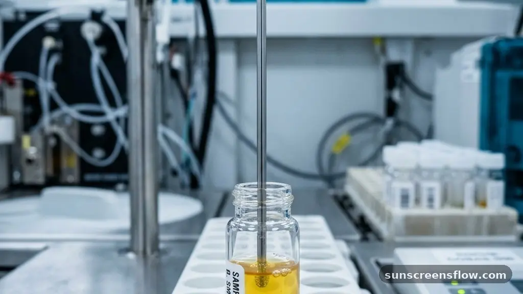
Because serum has already gone through the clotting process, it is devoid of fibrinogen, which means there is no risk of accidental clotting during the analysis process. This is particularly important for high-throughput machines that use narrow probes to aspirate samples; a tiny fibrin clot could cause a mechanical failure or lead to inaccurate readings.
However, plasma has its own distinct advantages. The primary benefit of plasma is speed. Because you do not have to wait for the blood to clot before centrifuging it, you can get results much faster.
In emergency departments or intensive care units where time is of the essence, plasma is the preferred choice. Additionally, plasma is often used for tests that measure specific hormones or volatile proteins that might be degraded during the slow process of clotting. By preventing the coagulation process, plasma preserves a more accurate representation of the patient’s metabolic state at the moment of the draw.
Comparing the Biochemical Profiles
To understand the difference, it helps to look at the protein composition. Both substances contain high concentrations of albumin and globulins, which are the primary proteins responsible for maintaining osmotic pressure and immune function.
However, the absence of fibrinogen in serum is the most significant biochemical difference. The following table provides a quick reference for these differences:
| Feature | Plasma | Serum |
|---|---|---|
| Clotting Factors | Present (e.g., Fibrinogen) | Absent (Consumed) |
| Preparation | Requires anticoagulant | Requires clotting time |
| Processing Time | Faster (no wait for clot) | Slower (requires clotting) |
| Primary Use | Emergency/Coagulation tests | Routine chemistry panels |
Beyond the proteins, the electrolyte levels can vary slightly between the two. During the clotting process, platelets release certain substances into the surrounding fluid.
For instance, the concentration of potassium in serum is often slightly higher than in plasma because potassium is released from cells during the clot formation process. This is a classic example of why lab technicians must be aware of the sample type: if they are measuring a patient’s potassium levels, they must know if the sample is serum or plasma to interpret the result correctly.
Addressing Common Topical Gaps
A common point of confusion is whether the color of the fluid differs between the two. Generally, both serum and plasma appear as a pale yellow or straw-colored liquid. However, variations in color can indicate underlying health issues.
A milky appearance (lipemia) suggests high levels of lipids, while a reddish tint (hemolysis) indicates that red blood cells have ruptured, potentially contaminating the sample. Hemolysis is a common problem in both serum and plasma, but it is often more pronounced in serum due to the longer time it spends in contact with the clot.
Another gap often missed is the impact of storage. Serum is generally more stable for long-term storage in biobanks. Because the clotting process has effectively “cleared” the sample of proteins that might otherwise trigger further reactions, serum samples are less likely to form precipitates over time.
Plasma, while useful for immediate testing, can occasionally form tiny fibrin clots during storage if the anticoagulant was not mixed perfectly or if the sample was subjected to freeze-thaw cycles. This makes serum the preferred medium for longitudinal studies and research where samples must be kept for months or years.
Clinical Significance and Future Directions
The distinction between these two fluids is more than just a classroom exercise; it is a fundamental pillar of modern medicine. Every time a physician orders a “CMP” (Comprehensive Metabolic Panel), they are likely looking at serum results.
When they order a “PT/INR” (Prothrombin Time/International Normalized Ratio), they are looking at plasma. This precision ensures that we can monitor everything from liver function and kidney health to the efficacy of blood-thinning medications.
As we move toward point-of-care testing and microfluidics, the ability to rapidly separate blood into its components will become even more important. New technologies are being developed that can perform plasma separation on a chip, allowing for near-instantaneous diagnostic results from a single drop of blood.
Despite these technological advancements, the fundamental physiological truth remains: the presence or absence of clotting factors defines the utility of the sample. Whether we are dealing with a routine cholesterol check or a complex analysis of clotting disorders, understanding the nature of the fluid we are analyzing is the first step toward accurate diagnosis and effective patient care.
Frequently Asked Questions
Is plasma the same as blood?
No, plasma is only one component of blood. Blood consists of cells (red blood cells, white blood cells, and platelets) suspended in plasma. Plasma is the liquid part that remains when all cellular components are removed, whereas whole blood includes both the cells and the liquid.
Why do doctors prefer serum for most routine tests?
Serum is preferred for many routine chemistry panels because it lacks fibrinogen. Fibrinogen can interfere with automated laboratory equipment, potentially clogging the sensitive probes used to measure substances like glucose, cholesterol, and electrolytes. Using serum ensures the sample remains stable and free of interference during the analysis process.
Can you turn plasma into serum?
No, you cannot turn plasma into serum. Once an anticoagulant has been added to blood to create plasma, the clotting process has been chemically inhibited. You cannot remove the anticoagulant and then induce the blood to clot in the same way it would have naturally, as the cellular integrity has already been compromised.
Does the centrifuge speed affect the quality of serum or plasma?
Yes, centrifuge speed and time are critical. If the centrifuge runs too slowly, the separation of cells from the liquid will be incomplete, leading to contamination. If it runs too fast or for too long, it can cause hemolysis, where red blood cells rupture and release their contents into the serum or plasma, potentially skewing test results.
Conclusion
In summary, the difference between serum and plasma is defined by the coagulation process. Plasma is the liquid fraction of blood collected with an anticoagulant, thereby preserving clotting factors like fibrinogen. Serum is the liquid fraction collected after the blood has been allowed to clot, resulting in a fluid that is free of these factors.
While both serve vital roles in diagnostic medicine, their specific compositions dictate their use: plasma is ideal for coagulation studies and emergency diagnostics where speed is paramount, while serum serves as the standard for routine chemistry panels due to its stability and lack of fibrinogen. By understanding these nuances, clinicians ensure that the diagnostic data they rely on is accurate, reproducible, and reflective of the patient’s true physiological state. Whether through the careful selection of collection tubes or the precise application of centrifugation, the management of these blood components remains a cornerstone of high-quality healthcare.