


When a healthcare professional draws a blood sample for analysis, the liquid portion that remains after processing is either serum or plasma. While these two components look nearly identical to the naked eye—typically appearing as a pale yellow, straw-colored fluid—their biochemical compositions are fundamentally different.
Understanding the serum vs plasma distinction is critical for laboratory technicians, clinicians, and researchers, as the choice between these two mediums can significantly influence the accuracy of diagnostic testing and the validity of clinical results. The primary difference lies in the presence or absence of coagulation factors, which dictates how the blood is handled during the initial stages of laboratory sample processing.
Defining the Fundamental Biological Composition
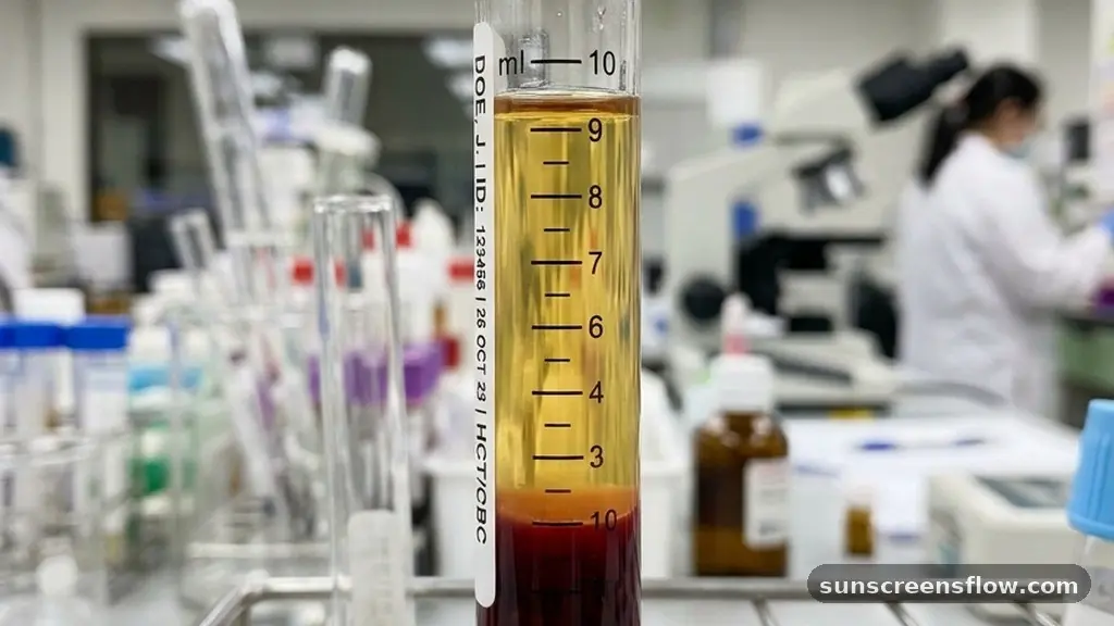
At the most basic level, blood is a complex tissue consisting of cells—red blood cells, white blood cells, and platelets—suspended in a liquid matrix. When this whole blood is collected, the way it is handled determines whether the end product is serum or plasma. Blood plasma composition includes water, salts, enzymes, antibodies, and other proteins, most notably albumin and globulins.
Crucially, plasma represents the liquid state of blood before it has had the opportunity to clot. Because it is obtained by preventing the clotting process, it retains all the soluble proteins that would otherwise be consumed during the formation of a clot.
In contrast, serum is essentially plasma that has been stripped of its clotting elements. When blood is allowed to clot, the clotting cascade is activated, leading to the conversion of soluble fibrinogen into an insoluble network of fibrin strands. This fibrin mesh traps blood cells and platelets, effectively removing them from the liquid fraction.
Once this process is complete, the remaining fluid—the supernatant—is what we identify as serum. Therefore, serum is defined as the liquid component of clotted blood. The absence of fibrinogen and other consumed factors is the hallmark that distinguishes serum from its plasma counterpart in a clinical setting.
The Role of Anticoagulants in Sample Preparation
To obtain plasma, medical professionals must intervene in the natural clotting process. This is achieved by adding an anticoagulant to the collection tube before the blood is introduced. Common anticoagulants include EDTA, sodium citrate, and heparin.
These substances work by chelating calcium ions or inhibiting specific enzymes within the coagulation pathway, thereby preventing the conversion of fibrinogen into fibrin. Because the blood is prevented from clotting, the resulting centrifuged blood fraction retains all the original proteins that were present in the circulating blood.
The selection of an anticoagulant is not arbitrary; it is dictated by the specific tests required. For instance, EDTA is the gold standard for hematology and complete blood counts because it preserves cell morphology exceptionally well. Sodium citrate is frequently used in coagulation studies because it is reversible, allowing for the precise timing of fibrin formation in a controlled environment. Heparin, on the other hand, is often preferred for certain chemistry panels because it does not interfere with electrolyte measurements as much as other agents might. Understanding which anticoagulant is present in a tube is vital because these chemicals can potentially interfere with downstream assays if the wrong tube type is selected for a specific analysis.
The Mechanics of Clotted Blood and Serum Extraction
Obtaining serum requires a different approach. Instead of using an anticoagulant, blood is collected into a plain tube or a serum separator tube (SST). In these tubes, the blood is allowed to sit undisturbed for a period, typically between 30 to 60 minutes, to facilitate natural coagulation.
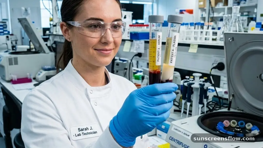
Once the clot has fully retracted, the sample undergoes blood sample centrifugation. The centrifugal force pushes the dense, clotted mass to the bottom of the tube, leaving the clear, straw-colored serum at the top.
The use of an SST has revolutionized modern phlebotomy. These tubes contain a specialized gel with a density intermediate between the serum and the clotted blood cells. During centrifugation, this gel moves upward to form a physical barrier between the serum and the clot.
This barrier prevents the serum from coming into contact with the cellular elements, which can otherwise release intracellular contents into the serum over time, leading to inaccurate test results. This streamlined method of laboratory sample processing ensures that the serum remains stable and uncontaminated, allowing for efficient transport and analysis in high-throughput clinical laboratories.
Clinical Chemistry and Diagnostic Testing Considerations
In the realm of clinical chemistry, the choice between serum and plasma often depends on the specific analyte being measured. Many standard chemistry panels, such as those measuring glucose, cholesterol, or liver enzymes, can be performed on either medium. However, there are notable exceptions.
For example, some assays measuring hormones or specific proteins may be sensitive to the presence of trace amounts of fibrinogen or other clotting factors found in plasma but absent in serum. Conversely, some proteins degrade rapidly in serum due to the activation of enzymes during the clotting process.
Furthermore, blood serum biomarkers are often preferred for certain immunological tests. Because serum is free from the interference of anticoagulants—which are essentially foreign chemical additives—it is frequently considered the “cleaner” sample for sensitive assays.
However, the time required for the blood to clot before centrifugation can be a significant drawback in emergency settings. In acute clinical scenarios where rapid turnaround time is essential, plasma is almost always the preferred choice because it can be processed and analyzed immediately after collection without the need for a long “clotting window.”
Comparing Serum and Plasma Properties
| Feature | Serum | Plasma |
|---|---|---|
| Coagulation Factors | Absent (consumed during clotting) | Present |
| Fibrinogen | Absent | Present |
| Preparation | Clotted blood, then centrifuged | Anticoagulated blood, then centrifuged |
| Processing Time | Slower (requires clotting time) | Faster (immediate centrifugation) |
| Additives | Usually none (or clot activator) | Requires anticoagulant (EDTA/Heparin) |
As shown in the table above, the primary differences are rooted in the processing methodology. The absence of fibrinogen in serum is the most significant biochemical distinction.
This protein is essential for the formation of a stable blood clot; once the clot is formed, the fibrinogen is no longer present in the liquid phase. In contrast, plasma acts as a reservoir for these factors, making it indispensable for coagulation studies where the integrity of the clotting cascade must be evaluated in real-time or under standardized laboratory conditions.
Addressing Topical Gaps: Why One Might Be Superior
A common question in the laboratory is whether one medium is objectively “better” than the other. The answer is highly contextual. Plasma is generally superior for speed and for tests that require the presence of clotting factors.
If a patient presents with a suspected clotting disorder, clinicians must use plasma because testing serum would provide zero information about the patient’s ability to form a clot—the very process that has already been artificially completed. Additionally, plasma is often preferred for measuring volatile substances that might be altered by the metabolic activity of cells during the clotting process.
Conversely, serum is often preferred for serological testing and the detection of antibodies. Because serum has been stripped of fibrinogen and other clotting proteins, it is less likely to cause interference in sensitive immunological assays. Some researchers argue that the clotting process itself can “clean” the sample, removing certain debris that might otherwise linger in plasma.
Ultimately, the “better” option is defined by the specific diagnostic goal. A laboratory director must balance the need for speed, the potential for chemical interference, and the specific requirements of the analytical instrumentation to determine the appropriate sample type for each test.
The Impact of Plasma Viscosity and Protein Stability
The protein composition of blood is dynamic, and both serum and plasma reflect this. Blood plasma proteins play a vital role in maintaining oncotic pressure and transporting hormones, lipids, and drugs throughout the body. Plasma viscosity is a clinical indicator that can reflect the total protein concentration, specifically the levels of immunoglobulins and fibrinogen. In conditions where these proteins are elevated, such as multiple myeloma or severe inflammation, the viscosity of the plasma increases, which can be measured as a diagnostic marker.
When comparing the stability of proteins in these two mediums, one must consider the degradation that occurs during the clotting process. The activation of the coagulation cascade involves a complex series of enzymatic reactions that can inadvertently activate or degrade other proteins present in the sample. For instance, certain cytokines or fragile peptides might be degraded by proteases released during clot formation.
In such cases, plasma is the superior medium because it is processed immediately, effectively “freezing” the protein composition at the moment of collection. This preservation is vital for research involving proteomics or the longitudinal study of biomarker stability in patients with chronic diseases.
Phlebotomy Best Practices and Sample Integrity
The journey of a blood sample begins with proper phlebotomy technique. Regardless of whether the goal is to obtain serum or plasma, the integrity of the sample depends on the skill of the collector.
Hemolysis—the rupture of red blood cells—is a common issue that can ruin both serum and plasma samples. When red blood cells break, they release hemoglobin and intracellular potassium into the surrounding liquid, which can lead to falsely elevated potassium results and interfere with spectrophotometric analysis.
To minimize the risk of hemolysis, phlebotomists must ensure that the needle size is appropriate, the tourniquet is not left on for too long, and the blood is gently mixed with the anticoagulant (if applicable) immediately after collection. Furthermore, the handling of the tube after collection is critical. For serum, the tube must be allowed to clot completely in a vertical position.
For plasma, the tube should be inverted gently multiple times to ensure the anticoagulant is thoroughly dispersed. Failure to follow these steps can lead to incomplete clotting (in the case of serum) or micro-clots (in the case of plasma), both of which can wreak havoc on automated laboratory instrumentation and lead to erroneous patient data.
Future Directions in Diagnostic Processing
As technology advances, the boundaries between traditional serum and plasma analysis continue to evolve. Automated systems are becoming increasingly sophisticated at handling various sample types, and the development of new, highly specific anticoagulants is reducing the potential for analytical interference. We are also seeing a shift toward point-of-care testing, where the rapid processing of plasma is becoming the standard for immediate decision-making in emergency departments and intensive care units.
Furthermore, the rise of “liquid biopsy” and the analysis of cell-free DNA (cfDNA) have placed a new emphasis on the quality of plasma samples. Because cfDNA is often present in very low concentrations, the presence of genomic DNA from white blood cells—which can leak into the sample if clotting or cell lysis occurs—is a major concern.
Consequently, specialized collection tubes designed to stabilize cell membranes and prevent the release of cellular contents are becoming standard in molecular diagnostics. Whether through the refinement of traditional methods or the adoption of new, stabilization-focused technologies, the goal remains the same: to ensure that the fluid analyzed in the laboratory is a true and accurate representation of the patient’s physiological state.
Frequently Asked Questions
Is there a visual way to distinguish between serum and plasma?
No, there is no reliable visual method to distinguish between the two. Both serum and plasma appear as pale yellow, straw-colored fluids. To identify them, you must know the type of collection tube used and whether an anticoagulant was present during the processing phase.
Why is fibrinogen absent in serum but present in plasma?
Fibrinogen is a soluble protein essential for clotting. During the preparation of serum, the blood is allowed to clot, which consumes the fibrinogen and converts it into an insoluble fibrin mesh. In plasma, an anticoagulant prevents this conversion, leaving the fibrinogen intact within the liquid.
Which sample type is better for emergency diagnostic testing?
Plasma is generally better for emergency testing because it can be processed and analyzed immediately after collection. Serum requires a mandatory waiting period for the blood to clot naturally, which adds significant time to the diagnostic workflow, making it less suitable for urgent clinical situations.
What role does the serum separator tube play in testing?
A serum separator tube (SST) contains a specialized gel that migrates during centrifugation to create a physical barrier between the serum and the clotted blood cells. This prevents the serum from interacting with the cells, which helps maintain sample stability and prevents contamination by intracellular components.
Conclusion
Distinguishing between serum vs plasma is more than a technicality; it is a fundamental aspect of clinical laboratory science that ensures the reliability of patient diagnostics. While both fluids originate from the same source, their divergent processing methods—specifically the use of anticoagulants versus the activation of the clotting cascade—create unique biochemical profiles suited for different types of testing. Plasma serves as a fast, comprehensive medium that retains its original protein composition, making it ideal for coagulation studies and rapid analysis.
Serum, by contrast, offers a clean, stable environment for many immunological and chemical assays, provided the clinician accounts for the proteins consumed during clot formation. Mastery of these differences, alongside rigorous adherence to phlebotomy standards and laboratory sample processing protocols, remains the cornerstone of high-quality healthcare. By selecting the correct medium for the specific diagnostic question at hand, medical professionals can guarantee the most accurate results, ultimately leading to better patient outcomes and more effective clinical decision-making.