

Blood is the life-sustaining fluid that circulates through our bodies, but once it is drawn for medical evaluation, it becomes a complex mixture of cells and liquid components. When you look at a test tube of blood, the difference between serum and plasma—is the fundamental starting point for any laboratory analysis.
While both appear as pale, straw-colored fluids, their biochemical compositions are distinct due to the presence or absence of specific proteins involved in the clotting cascade. Understanding the divergence between these two substances is essential for clinicians, phlebotomists, and patients alike, as the choice between them can significantly influence the accuracy of diagnostic testing and the reliability of clinical results.
The Biological Foundations of Blood Components
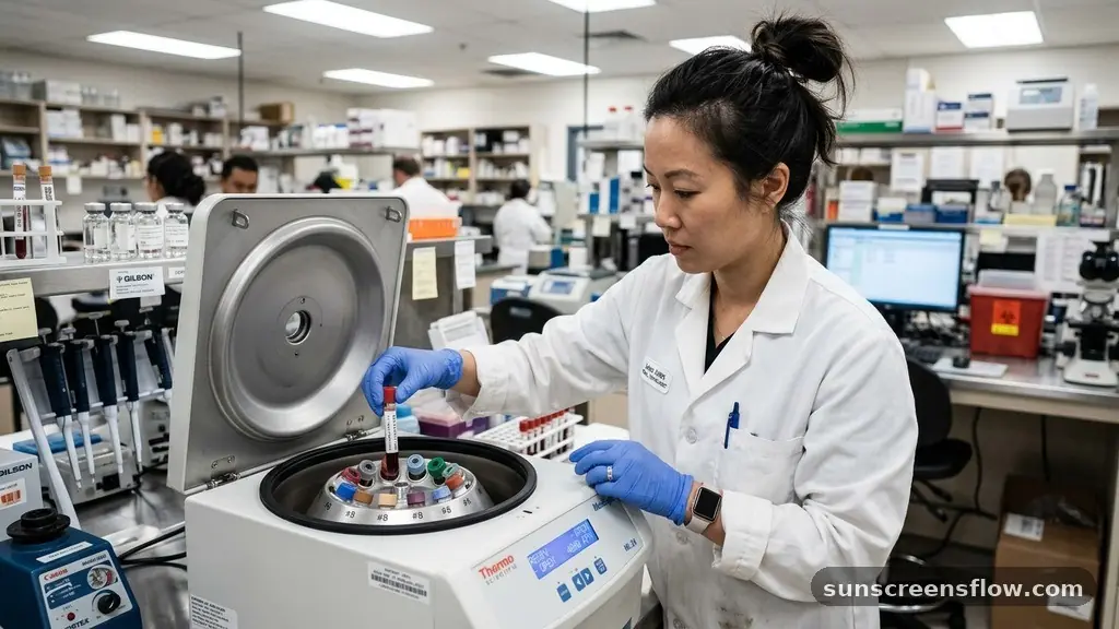
At its most basic level, whole blood consists of formed elements—red blood cells, white blood cells, and platelets—suspended in a liquid matrix. To isolate the liquid portion, laboratories utilize centrifugation, a process that spins blood at high speeds to separate components based on density. However, the resulting liquid depends entirely on how the blood was handled before and during this spinning process.
If the blood is allowed to clot, the resulting fluid is serum. If the blood is prevented from clotting through the addition of chemical agents, the resulting fluid is plasma.
This difference is not merely academic; it is rooted in the physiological process of homeostasis. When blood vessels are injured, the body initiates a series of chemical reactions to prevent excessive bleeding. This process, known as blood coagulation, involves the rapid conversion of soluble proteins into an insoluble mesh.
By manipulating this process in a clinical setting, laboratory professionals can extract specific components that are optimized for different types of assays. Because serum is the byproduct of completed coagulation, it is inherently depleted of certain proteins that were consumed during the formation of the clot. Conversely, plasma retains these proteins, providing a more “authentic” snapshot of the circulating blood chemistry at the moment of the draw.
Defining Blood Plasma and its Composition
Blood plasma is the liquid, cell-free part of blood that has been treated with anticoagulants to prevent the coagulation process from occurring. When blood is collected in a tube containing an anticoagulant like EDTA, heparin, or sodium citrate, the blood cells are separated, but the liquid remains chemically similar to the blood as it exists inside your veins. Because the clotting factors, particularly fibrinogen, have not been consumed or converted into a solid mass, they remain suspended within the plasma.
This makes plasma the preferred medium for tests that require an accurate measurement of proteins that are otherwise depleted in serum. For example, in coagulation studies, clinicians must use plasma because the very factors they are trying to measure—such as prothrombin time or activated partial thromboplastin time—would be absent or significantly altered in a serum sample.
Plasma is approximately 90% water, with the remaining 10% consisting of dissolved solutes, including glucose, mineral ions, hormones, carbon dioxide, and various serum proteins like albumin and globulins. Because plasma is obtained immediately after centrifugation without waiting for a clot to form, it is often the faster choice for urgent medical situations where time-sensitive data is required to make life-saving decisions.
The Formation and Characteristics of Serum
Serum is defined as the liquid portion of blood that remains after the blood has been allowed to clot. Unlike plasma, serum does not contain fibrinogen or other clotting factors because these components were used up during the formation of the clot.
The process of obtaining serum involves collecting blood in a plain tube (often with no additive or a clot activator) and allowing it to sit undisturbed for a period, typically 30 to 60 minutes. As the blood sits, the clotting cascade runs its course, trapping red and white blood cells in a fibrin mesh.
Once the clot has formed, the tube is subjected to centrifugation. The dense clot sinks to the bottom, while the clear, straw-colored liquid—the serum—remains as the supernatant at the top. This fluid is rich in antibodies, electrolytes, and enzymes, making it ideal for a wide range of biochemical assays and serological testing.
Because serum lacks the proteins that create clots, it is much less likely to cause interference in certain automated testing instruments. Modern phlebotomy often utilizes a serum separator tube (SST), which contains a gel barrier that moves between the serum and the clot during spinning, ensuring that the serum can be easily extracted without contamination from the cellular layer.
Key Differences in Laboratory Processing
The primary operational difference between these two samples lies in the blood draw tubes and the preparation time required. When a phlebotomist collects blood, the color of the tube cap indicates the type of anticoagulant present, which dictates whether the lab will ultimately process the sample into plasma or serum.
| Feature | Plasma | Serum |
|---|---|---|
| Clotting Factors | Present (e.g., Fibrinogen) | Absent (Consumed) |
| Anticoagulant Use | Required (EDTA, Heparin, etc.) | None (or Clot Activator) |
| Processing Time | Immediate | Requires Clotting Time |
| Primary Use | Coagulation, Emergency Tests | Chemistry, Immunology |
As shown in the table, the inclusion of an anticoagulant is the deciding factor in the workflow. Using an anticoagulant like heparin allows for immediate processing, which is critical in emergency departments where a delay of 30 minutes to allow for clotting could be detrimental to patient care.
However, for routine metabolic panels or lipid profiles, serum is frequently the gold standard. The choice is often dictated by the specific requirements of the diagnostic equipment being used, as some reagents react poorly to the presence of anticoagulants, which could lead to false results in a clinical pathology report.
The Role of the Buffy Coat in Separation
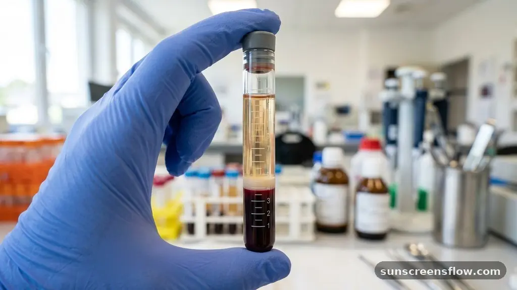
When observing a centrifuged sample, you will notice distinct layers. The bottom layer is composed of red blood cells, which are the densest. The top layer is the liquid (either plasma or serum).
Between these two layers lies a thin, often whitish or grayish transition zone known as the buffy coat. This layer contains the majority of the white blood cells and platelets.
In the context of laboratory analysis, the presence of the buffy coat is more pronounced in plasma samples because the cells have not been trapped in a dense clot. When preparing plasma, technicians must be careful to aspirate only the clear liquid above the buffy coat to avoid cellular contamination.
In serum production, the clot formation process is more chaotic, and while the buffy coat is technically present within the clot, it is not as clearly demarcated for manual removal. Understanding the composition of the buffy coat is vital for researchers performing genomic studies or specialized white blood cell counts, as this layer is the concentrated source of DNA-containing cells within the blood sample.
Clinical Applications and Diagnostic Utility
The decision to use serum versus plasma is driven by the specific diagnostic goal. Serum proteins are the cornerstone of many immune system assessments. For example, when measuring antibody titers or performing an electrophoresis to check for protein disorders, serum is almost always preferred because the absence of fibrinogen reduces the overall protein complexity of the sample, allowing for a cleaner reading of the specific immunoglobulins present.
On the other hand, plasma is indispensable for monitoring the status of the coagulation system. If a patient is on blood-thinning medication, such as warfarin or heparin, their plasma must be tested regularly to ensure that the clotting factors are within a therapeutic range.
If serum were used, the lab would be unable to measure the effectiveness of the drug because the very factors being influenced by the medication would have been removed during the clotting process. Furthermore, plasma is the preferred sample for many molecular tests, including viral load testing for HIV or Hepatitis C, as the preservation of the sample’s original state ensures that viral particles are not trapped within a clot or degraded by the coagulation process.
Addressing Topical Gaps: Why Serum Sometimes Beats Plasma
While plasma is faster to process, there are valid clinical reasons why serum remains the preferred sample for many routine tests. One of the most significant issues with plasma is “fibrin interference.” If a plasma sample is not processed perfectly, small strands of fibrin can remain in the liquid.
These strands can clog the delicate probes of automated chemistry analyzers, leading to instrument downtime and potentially erroneous results. Because serum has already undergone the full clotting process, there is no risk of residual fibrin formation during the subsequent testing phase.
Additionally, some chemical assays are sensitive to the specific anticoagulants used to create plasma. For instance, EDTA is a chelating agent that binds to calcium. If a test requires the measurement of calcium levels, using an EDTA-plasma sample would result in an artificially low reading because the anticoagulant has sequestered all the calcium ions in the tube.
In such cases, serum is the only viable option for accurate laboratory analysis. By utilizing serum, the clinician ensures that the chemical environment of the sample remains as close to the natural state as possible, without the interference of exogenous chemicals.
The Importance of Proper Phlebotomy and Sample Handling
The quality of the final result is only as good as the quality of the sample collected. Phlebotomy is not just about drawing blood; it is about maintaining the integrity of the specimen until it reaches the analyzer. Improper mixing of blood with anticoagulants can lead to micro-clots in plasma samples, which can cause significant errors in hematology and chemistry results. Conversely, failing to allow sufficient clotting time for serum samples can lead to a “hemolyzed” or incomplete clot, which complicates the separation process.
Proper handling also involves managing the temperature and transport of the tubes. Some analytes are sensitive to heat and will degrade if the sample is left sitting in a warm environment. Others may require immediate cooling to prevent the cells from leaking their contents into the liquid, a process known as hemolysis.
When blood cells rupture, they release intracellular components like potassium into the serum or plasma, which can cause a false elevation in test results. This is why standard operating procedures in clinical pathology emphasize the importance of gentle inversion of tubes and prompt transport to the laboratory.
Common Challenges in Modern Hematology
Despite advancements in technology, laboratories still face challenges when differentiating and processing these samples. One major hurdle is the presence of lipemia, or high levels of fat in the blood, which can make both serum and plasma appear cloudy or milky.
This cloudiness can interfere with light-based diagnostic tests, making it difficult for the machine to “see” through the sample. In such cases, the laboratory may need to perform high-speed centrifugation or use specific reagents to clear the sample before an accurate reading can be obtained.
Another challenge is the variability in patient physiology. Patients with certain blood disorders may have abnormally high or low levels of clotting factors, which can affect the consistency of the clot in serum tubes or the stability of plasma samples. Furthermore, the rise of point-of-care testing has introduced a new dynamic where blood is often tested at the bedside.
In these settings, the distinction between serum and plasma is often handled by the device itself, using microfluidic cartridges that perform the separation automatically. As we move toward more personalized medicine, the ability to rapidly and accurately distinguish between these two components will continue to be a vital skill for healthcare professionals.
Frequently Asked Questions
Is serum the same thing as plasma?
No, they are not the same. While both are the liquid components of blood, serum is the fluid remaining after blood has clotted, meaning it lacks clotting factors like fibrinogen. Plasma is the liquid obtained when an anticoagulant is added to prevent clotting, so it retains these factors.
Which is better for diagnostic testing, serum or plasma?
Neither is objectively “better”; it depends on the test. Serum is preferred for chemistry and immunology tests where clotting factors might interfere with the results. Plasma is essential for coagulation studies and emergency tests where speed is critical and the presence of clotting factors is required.
Why would a doctor order a plasma test instead of a serum test?
A doctor orders a plasma test when they need to evaluate the coagulation system or require a rapid turnaround time. Because plasma does not require a waiting period for clotting, it is the standard choice for urgent diagnostic testing and for monitoring patients on anticoagulant medications.
Can I use serum for a coagulation study?
No, you cannot use serum for a coagulation study. Coagulation tests are specifically designed to measure how well the blood’s clotting factors function. Since these factors are consumed during the clotting process used to create serum, a serum sample would provide inaccurate or non-existent results for these tests.
Conclusion
Distinguishing between serum and plasma is a fundamental aspect of modern medicine. While both substances share a common origin and look nearly identical to the naked eye, their biochemical differences—specifically the presence or absence of clotting factors—dictate their clinical utility. Serum, obtained after the completion of blood coagulation, provides a stable medium for a vast array of immunological and metabolic tests.
Plasma, preserved by anticoagulants like EDTA or heparin, offers a faster, more representative sample for emergency diagnostics and coagulation studies. By understanding the nuances of centrifugation, the role of the buffy coat, and the potential for analytical interference, laboratory professionals ensure the highest standards of diagnostic testing. Whether in a routine check-up or a critical care scenario, the careful separation of these two fluids remains a silent but essential pillar of patient care and clinical pathology.