


Blood is the life-sustaining fluid that circulates through our cardiovascular system, but to a clinical laboratory scientist, it is a complex cocktail of cells, proteins, and electrolytes. When a sample of blood is drawn for analysis, the liquid portion that remains after the cellular components are removed is either plasma or serum. Understanding what’s the difference between plasma and serum is fundamental to modern medicine, as the choice between these two fluids dictates which diagnostic tests can be performed and how the sample must be handled from the moment of collection to the final analysis in the laboratory.
The Fundamental Composition of Blood
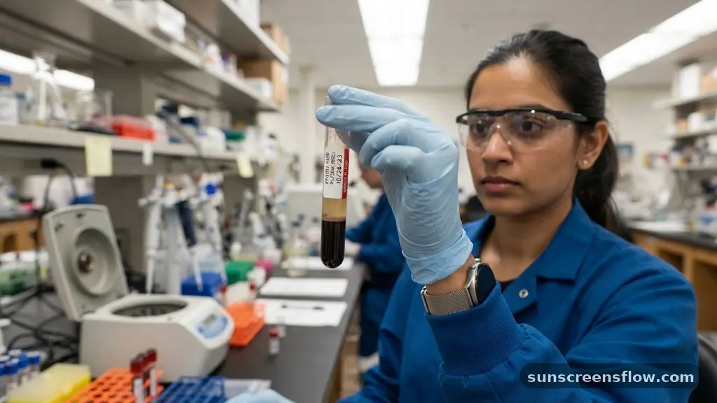
To grasp the distinction between these two liquids, one must first understand the architecture of whole blood. Whole blood consists of formed elements—red blood cells, white blood cells, and platelets—suspended in a liquid matrix known as plasma.
Plasma represents approximately 55% of the total blood volume. It is a straw-colored, protein-rich fluid that carries hormones, nutrients, waste products, and, crucially, the proteins involved in blood coagulation.
When blood remains in its natural state within the vascular system, it stays liquid. However, once blood is extracted, the body’s innate defense mechanism triggers the clotting cascade. This process involves the conversion of the soluble protein fibrinogen into an insoluble mesh of fibrin, which traps blood cells to form a clot.
Plasma, by definition, contains all of the original clotting factors because it is isolated from the blood before this cascade completes. Serum, on the other hand, is the fluid that remains after the blood has been allowed to clot. Therefore, the primary physiological difference lies in the presence or absence of these specialized coagulation proteins.
Laboratory Collection and Processing Methods
The pathway to obtaining either fluid begins with the specific collection requirements set by the laboratory. If a clinician requires a plasma sample, they must use a collection tube that contains an additive known as an anticoagulant. These chemical agents prevent the clotting cascade from ever initiating, ensuring that the liquid portion retains its full complement of clotting factors.
Common anticoagulants include EDTA, heparin, and sodium citrate. Each of these works through a different mechanism—for instance, EDTA chelates calcium, which is a necessary cofactor for the clotting process, while heparin activates antithrombin III to inhibit thrombin and factor Xa.
To obtain serum, the phlebotomist uses a “clot activator” tube, often identified by a red or gold cap. The blood is allowed to sit undisturbed for a period, usually between 30 to 60 minutes, to allow the natural coagulation process to finish. Once the clot has formed, the sample undergoes centrifugation.
This process uses high-speed rotation to separate the solid clot and cells from the liquid supernatant, which is the serum. In contrast, plasma is also separated via centrifugation, but because the anticoagulants have already done their work, the separation occurs without the formation of a solid fibrin clot. This procedural difference is the defining factor in laboratory workflows.
The Role of Clotting Factors and Proteins
The presence of prothrombin and other various clotting factors is the hallmark of plasma. In the body, these proteins exist in a dormant state, ready to be activated at the site of a vascular injury.
Because plasma is collected in the presence of an agent that halts coagulation, these proteins remain present in the final sample. This makes plasma an essential medium for tests that specifically measure the activity of the coagulation system, such as Prothrombin Time (PT) or Activated Partial Thromboplastin Time (APTT).
Serum, by definition, lacks these factors. During the formation of the serum, the fibrinogen is consumed and converted into the fibrin mesh that constitutes the clot. Consequently, serum cannot be used for coagulation studies.
However, the absence of these proteins is actually an advantage for certain types of chemical analysis. Because the clotting process has already occurred, there is no risk of the sample spontaneously clotting inside the delicate tubing of an automated clinical analyzer, which could cause instrument downtime or erroneous results. Scientists must carefully select their medium based on whether they need to measure the coagulation system itself or the underlying chemical profile of the patient’s blood.
Visual and Chemical Comparison
When assessing the samples side-by-side, professionals often look for specific physical indicators. While both fluids are yellowish, there are subtle differences in their appearance and consistency. Plasma can sometimes appear slightly more opaque or cloudy if the centrifugation process was not sufficient to remove all platelets or if the patient has a high lipid content. Serum, having undergone the clotting process, is generally clearer because the removal of the fibrinogen and the retraction of the clot tend to leave a cleaner liquid layer.
Chemically, the two fluids are remarkably similar in terms of electrolyte levels, such as sodium, potassium, and chloride. However, there are minor variations. For instance, the concentration of certain proteins may differ slightly because the clotting process releases specific substances from the platelets during clot formation.
Potassium levels, for example, can be slightly higher in serum than in plasma because potassium is released from platelets as they break down during the clotting process. This is a critical consideration for clinicians interpreting lab results; if a test is extremely sensitive to potassium levels, the choice of anticoagulant or the decision to use serum over plasma can lead to different clinical interpretations of a patient’s health status.
Clinical Applications and Diagnostic Utility
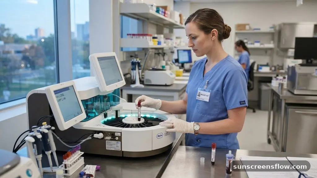
The utility of these fluids spans the entire breadth of medical diagnostics. Plasma is the preferred sample for specialized assays that require the presence of all clotting factors.
Furthermore, because plasma can be processed almost immediately after collection—without waiting for the blood to clot—it is the standard choice for emergency or “stat” testing where turnaround time is critical. It is also the sample of choice for molecular diagnostics, such as PCR testing for viral loads, where the integrity of the sample must be preserved exactly as it existed in the patient’s body.
Serum is the workhorse of routine chemistry panels. Most automated analyzers are calibrated specifically for serum, as it is a very stable medium for long-term storage and high-throughput analysis. Typical tests performed on serum include:
- Electrolyte panels to assess hydration and kidney function.
- Liver function tests to measure enzyme levels like ALT and AST.
- Lipid profiles to evaluate cholesterol and triglyceride levels.
- Serological testing for antibodies, which is crucial for identifying immune responses to infections or autoimmune conditions.
Because serum is devoid of fibrinogen, it is less likely to interfere with optical measurement techniques used in these automated systems, making it the preferred choice for clinical chemistry.
Comparing Sample Characteristics
| Feature | Plasma | Serum |
|---|---|---|
| Clotting Factors | Present | Absent |
| Fibrinogen | Present | Absent |
| Preparation Time | Immediate (after spin) | 30-60 minutes (clotting) |
| Anticoagulants | Required | Not used |
| Primary Use | Coagulation studies, emergency tests | Chemistry, serology, immunology |
This table highlights the fundamental differences that laboratory staff must consider. The requirement for an anticoagulant in plasma collection is perhaps the most significant operational difference.
If a technician accidentally places a sample intended for a coagulation test into a plain red-top tube, the sample will clot, and the test will be invalidated. Conversely, using a lavender-top tube (which contains EDTA) for a test that requires a serum matrix can lead to skewed results, particularly for tests like calcium, which the EDTA would bind and artificially lower.
Addressing Topical Gaps: Stability and Storage
One common question that arises in clinical settings is whether plasma or serum is more stable for long-term storage in a biobank. Generally, serum is considered the more stable medium for long-term storage because the absence of fibrinogen prevents the formation of “fibrin strands” that can develop in plasma samples over time, especially after repeated freeze-thaw cycles. These strands can interfere with subsequent analysis, potentially clogging the probes of diagnostic instruments.
However, plasma offers the advantage of higher yield. Because the clot does not form, the volume of liquid recovered from the blood is higher. In pediatric or geriatric patients where blood collection volume is strictly limited, plasma is often preferred simply because it provides more material for the lab to work with.
Furthermore, for certain hormones and unstable proteins, the presence of specific anticoagulants in plasma can help prevent degradation that might occur during the slower clotting process required for serum. Understanding these nuances allows laboratory directors to optimize their protocols for accuracy, efficiency, and patient safety, ensuring that every sample provides the most reliable data possible.
The Impact of Anticoagulants on Test Accuracy
The choice of anticoagulant is not merely a procedural preference; it is a chemical necessity that directly impacts the validity of diagnostic results. EDTA is excellent for hematology because it preserves cell morphology, but it is a poor choice for tests that measure calcium or iron, as it binds these ions tightly, yielding a falsely low result. Heparin, in contrast, is often used for chemistry panels because it does not affect most electrolyte levels, yet it can interfere with certain PCR-based assays by inhibiting the enzymes required for DNA amplification.
This interaction between the sample additive and the analytical method is why laboratory information systems are programmed with strict “order of draw” and “tube type” requirements. If a nurse draws a blood sample and places it in the wrong tube, the chemical interference caused by the wrong anticoagulant could lead to a misdiagnosis.
For example, a patient might appear to have dangerously low calcium levels simply because the sample was collected in an EDTA tube rather than a serum separator tube. These technical details are the silent safeguards of modern medicine, preventing analytical errors that could have significant clinical consequences for the patient.
Ensuring Sample Integrity in Modern Diagnostics
In the modern era of high-speed clinical diagnostics, the speed and efficiency of sample processing remain paramount. The move toward “point-of-care” testing has further emphasized the need to understand these fluid differences.
Devices that can analyze a drop of blood at the bedside often rely on internal mechanisms to mimic either plasma or serum isolation. By understanding the underlying physics and chemistry—the role of fibrinogen, the effect of centrifugation, and the impact of anticoagulants—clinicians are better equipped to troubleshoot results that seem inconsistent with the patient’s clinical presentation.
Ultimately, both plasma and serum are indispensable tools. One is the fluid of the active, circulating system, captured in its dynamic state; the other is the fluid of the post-coagulation environment, stripped of its clotting potential to provide a clean, stable window into the body’s metabolic state.
As diagnostic technology evolves, the ability to discern the subtle differences between these two will remain a cornerstone of professional medical practice. Whether it is identifying an antibody in a serum sample or monitoring a clotting factor in plasma, the precision of our diagnostic capabilities rests on this fundamental biological distinction.
FAQ: Common Questions About Plasma and Serum
Can plasma be converted into serum?
No, plasma cannot be converted into serum. Serum is defined by the absence of clotting factors that are consumed during the coagulation process. Once these factors are removed or consumed, they cannot be restored, and the fundamental chemical composition of the fluid is permanently altered.
Why is serum often preferred for chemistry panels?
Serum is preferred because it is free of fibrinogen and other clotting proteins, which can interfere with the optical systems of automated chemistry analyzers. Additionally, serum is generally more stable for long-term storage and less prone to the formation of fibrin strands that can clog diagnostic instrumentation.
What is the main visual difference between the two?
While both are yellowish, serum is typically clearer because the clotting process has removed fibrinogen. Plasma may appear slightly more opaque or cloudy, particularly if the centrifugation was incomplete or if the patient has high levels of lipids or other suspended particles in their blood.
Does the choice of anticoagulant affect the test results?
Yes, significantly. Anticoagulants like EDTA can bind to minerals like calcium or iron, leading to falsely low results for those analytes. Similarly, heparin can interfere with molecular tests like PCR. Using the incorrect tube type for a specific test can directly compromise the accuracy of the diagnostic data.
Conclusion
Distinguishing between plasma and serum is more than just a technical exercise; it is a critical component of diagnostic accuracy. Plasma, obtained by preventing coagulation, provides a snapshot of the blood in its circulating state, complete with all clotting factors, making it essential for coagulation and molecular studies. Serum, obtained by allowing the blood to clot, provides a stable, clean matrix ideal for routine chemistry and immunology.
By understanding the distinct processing requirements, such as the use of anticoagulants like EDTA or heparin versus the clot-activation process, healthcare professionals ensure that the right diagnostic test is performed on the right medium. This foundation of knowledge supports the reliability of clinical data, ultimately leading to better patient outcomes and more precise medical decision-making in the laboratory and beyond.